VARICOCELE

A varicocele is an enlargement of the veins that transport oxygen-depleted blood away from the testicle. A varicocele is an enlargement of the veins within the loose bag of skin that holds your testicles (scrotum). A varicocele is similar to a varicose vein you might see in your leg.

Varicocele treatment might not be necessary. Many men with varicoceles are able to father a child without any treatment. However, if your varicocele causes pain, testicular atrophy or infertility or if you are considering assisted reproductive techniques, you might want to undergo varicocele repair. 

The purpose of surgery is to seal off the affected vein to redirect the blood flow into normal veins. In cases of male infertility, treatment of a varicocele might improve or cure the infertility or improve the quality of sperm if techniques such as in vitro fertilization (IVF) are to be used.

Clear indications to repair a varicocele in adolescence include progressive testicular atrophy, pain or abnormal semen analysis results. Although treatment of a varicocele generally improves sperm characteristics, it's not clear if an untreated varicocele leads to progressive worsening of sperm quality over time.


RISKS

These are the commoner risks. There may be other unusual risks that have not been listed here. Please ask you surgeon if you have any general or specific concerns.There are risks associated with any anaesthetic.You may have side effects from any drugs used. The commoner side effects include light-headedness, nausea, skin rash and constipation.

The correction of a Varicocele has very few risks but can include:

  • Build-up of fluid around the testicles (hydrocele)
  • Recurrence of varicoceles
  • Infection
  • Damage to an artery

Some of the above risks are more likely if you smoke, are overweight, diabetic, have high blood pressure or have had previous heart disease.

Method of repair used: Open surgery. 

This treatment usually is done during general anaesthetic. Commonly, your surgeon will approach the vein through your groin (inguinal or subinguinal), but it's also possible to make an incision in your abdomen or below your groin.

Advances in varicocele repair have led to a reduction of post-surgical complications. One advance is the use of the surgical microscope, which enables the surgeon to see the treatment area better during surgery. 

Another is the use of Doppler ultrasound, which helps guide the procedure.

You might be able to return to normal, nonstrenuous activities after two days. As long as you're not uncomfortable, you might return to more strenuous activity, such as exercising, after two weeks.

Pain from this surgery generally is mild but might continue for several days or weeks. Your doctor might prescribe pain medication for a limited period after surgery. After that, your doctor might advise you to take over-the-counter painkillers, to relieve discomfort.

Your doctor might advise you not to have sex for a period of time. Most often, it will take several months after surgery before improvements in sperm quality can be seen with a semen analysis. This is because it takes approximately three months for new sperm to develop.

Open surgery using a microscope and subinguinal approach (microsurgical subinguinal varicocelectomy) has the highest success rates when compared with other surgical methods.

When you are discharged, take care of the following:

  • Take it easy for the first 2 days after the procedure to minimise the risk of bleeding and bruising
  • Wear firm-fitting underwear to help minimize bleeding
  • You can remove your dressing/plaster after 2 days
  • Sexual activity can be resumed after 1 week if you are comfortable
  • Finish all prescribed medication
  • Contact your doctor if you have severe pain, bleeding or fever

Torsion of Testis

Testicular torsion occurs when the testicle 
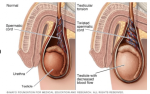
rotates on the spermatic cord, which brings blood to the testicle from the abdomen. If the testicle rotates several times, blood flow to it can be entirely blocked, causing damage more quickly.

It's not clear why testicular torsion occurs. Most males who get testicular torsion have an inherited trait that allows the testicle to rotate freely inside the scrotum. This inherited condition often affects both testicles. But not every male with the trait will have testicular torsion.

Testicular torsion often occurs several hours after vigorous activity, after a minor injury to the testicles or while sleeping. Cold temperature or rapid growth of the testicle
during puberty also might play a role.

Testicular torsion usually requires emergency surgery. If treated quickly, the testicle can usually be saved. But when blood flow has been cut off for too long, a testicle might become so badly damaged that it has to be removed.
RISKS

Age. Testicular torsion is most common between ages 12 and 18.
Previous testicular torsion. If you've had testicular pain that went away without treatment (intermittent torsion and detorsion), it's likely to occur again. The more frequent the bouts of pain, the higher the risk of testicular damage.
Family history of testicular torsion. The condition can run in families.

A Testis Torsion can cause the following complications:

Damage to or death of the testicle. When testicular torsion is not treated for several hours, blocked blood flow can cause permanent damage to the testicle. If the testicle is badly damaged, it has to be surgically removed.
Inability to father children. In some cases, damage or loss of a testicle affects a man's ability to father children.

When you are discharged, take care of the following:

Take it easy for the first 2 days after the procedure to minimise the risk of bleeding and bruising
Use scrotal support for 10 days
Keep the dressing on for 7 days
The stitches will dissolve by itself
Call the rooms if you experience bleeding, swelling, fever, pain or warm/red scrotum

BCG Instillations

BCG instillations

Bacillus Calmette-Guerin (BCG) is the main  intravesical immunotherapy for early stage bladder cancer.

It is made from a weakened strain of Mycobacterium bovis, a vaccine for tuberculosis.Immunotherapy is used to prompt the immune system into attacking cancer cells. BCG is a liquid drug that can be deposited directly into your bladder through a catheter. Doctors have been using this method of treating superficial bladder cancer for 40 years.BCG is appropriate for non-invasive (stage 0) and minimally invasive (stage 1) bladder cancers. It usually follows a procedure called 

transurethral resection of bladder tumor (TURBT).

It is intended to help prevent recurrence. This treatment only affects cells

inside the bladder. It’s not useful for later stage bladder cancer that has

spread into or beyond the bladder lining, or to other tissues and organs.


Is there any preparation involved?

It is important that you follow your doctor’s instructions for what to do before and after the procedure. Tell your doctor about all the medications you take. Certain immunosuppressants, antimicrobial therapies, and radiation therapies can interfere with BCG treatment.

You will be advised to limit your fluid intake for four hours prior to the procedure. You might be told to avoid caffeine for a few hours longer than that, because it is a diuretic and could make things more difficult.

You’ll be asked to urinate just before the procedure, so you’ll be able to hold the medication in your bladder for several hours.

What happens during treatment?

A urinary catheter is inserted through your urethra and into your bladder. Then the BCG solution is injected into the catheter. The catheter is clamped off so the solution stays in your bladder. Some doctors may remove the catheter at this time.

You have to hold the medicine in your bladder. You’ll be instructed to lie on your back and to roll from side to side to make sure the solution reaches your entire bladder.

After about two hours, the catheter is unclamped so the fluid can be drained. If the catheter was already removed, you’ll be asked to empty your bladder at this time.

What can I expect following treatment?

For six hours after each treatment, you’ll have to be very careful when you urinate to avoid transmitting BCG to others. Men should urinate while seated to avoid splashing.

Disinfect the urine by adding 2 cups of bleach into the toilet. Let it stand for about 20 minutes before flushing. You should also wash your genital area very carefully after you urinate, so your skin doesn’t become irritated from the BCG. Wash your hands thoroughly, too.

Men can pass BCG to their partner during sex. For that reason, you should avoid sex for 48 hours after each treatment. Use a condom between treatments and for six weeks following your final treatment.

Women should avoid getting pregnant or breastfeeding while on BCG therapy.

Treatment is usually given every week for six weeks.

What side effects can occur?

One benefit of BCG is that while it affects the cells in your bladder, it doesn’t have a major effect on any other part of your body. But there can be a few side effects such as:

Contact your doctor if you experience one of the above symptoms, have increasing pain or unable to urinate.

Kidney Stone manipulation: LASER

Kidney Stone manipulation: LASER

Kidney stones form in your kidneys. As stones move into your ureters (the thin tubes that allow urine to pass from your kidneys to your bladder) signs and symptoms can result. Signs and symptoms of kidney stones can include severe pain, nausea, vomiting, fever, chills and blood in your urine.

Laser lithotripsy is a procedure to break apart kidney stones in the urinary tract. It is done with a ureteroscope passed into the tubes of the urinary tract. Incisions are not needed.

The laser breaks the kidney stones into smaller pieces that can either be removed by the surgeon or pass out of the body in the urine.

A ureteroscope is an instrument with a long thin tube that is hollow like a straw. The doctor can use this scope to view the urinary tract, find kidney stones, and pass instruments to the stone.

The scope enters the urinary tract through the urethra, the tube that carries urine out of the body. The scope continues to pass through the urethra, bladder, and into the ureter or kidney (if necessary) to access the stone.

Once the doctor sees the stone, a fiber will be sent through the scope to the stone. The fiber can create a laser beam to break up the stone. Small pieces may be removed using a basket that is passed through the scope. Small sand-like pieces may remain and will be gradually passed through the urine.

A temporary stent may be placed in the ureter. The stent will keep the ureter open, improve urine flow, and help the stone pieces pass. The stent will be removed after a few days or weeks.

RISKS

These are the commoner risks. There may be other

unusual risks that have not been listed here. Please

ask you surgeon if you have any general or specific

concerns. There are risks associated with any anaesthetic.

You may have side effects from any drugs used. The

Commoner side effects include light-headedness,

nausea, skin rash and constipation.


Factors that increase your risk of developing kidney stones include:

  • Family or personal history. If someone in your family has kidney stones, you're more likely to develop stones, too. And if you've already had one or more kidney stones, you're at increased risk of developing another.
  • Dehydration. Not drinking enough water each day can increase your risk of kidney stones. People who live in warm climates and those who sweat a lot may be at higher risk than others.
  • Certain diets. Eating a diet that's high in protein, sodium (salt) and sugar may increase your risk of some types of kidney stones. This is especially true with a high-sodium diet. Too much salt in your diet increases the amount of calcium your kidneys must filter and significantly increases your risk of kidney stones.
  • Being obese. High body mass index (BMI), large waist size and weight gain have been linked to an increased risk of kidney stones.
  • Digestive diseases and surgery. Gastric bypass surgery, inflammatory bowel disease or chronic diarrhoea can cause changes in the digestive process that affect your absorption of calcium and water, increasing the levels of stone-forming substances in your urine.
  • Other medical conditions. Diseases and conditions that may increase your risk of kidney stones include renal tubular acidosis, cystinuria, hyperparathyroidism, certain medications and some urinary tract infections.

Some of the above risks are more likely if you smoke, are overweight, diabetic, have high blood pressure or have had previous heart disease.

When you are discharged, take care of the following:

  • You can continue with your normal daily activities (eg. Driving, sexual activities, walking, exercise & travel)
  • Drink enough fluids (1-2 litres per day)
  • Take your medicine as prescribed
  • You can relax in a hot bath or shower if you experience discomfort
  • Rest when you can
  • If urine becomes blood stained, increase your fluid intake
  • Make sure you attend to your follow-up appointment to remove the JJ-stent: usually within 2 weeks of stent placement if removed in theatre. +/- 2 day’s if to be removed in the rooms (your doctor will confirm whether it will be taken out in theatre or in the rooms)
  • Contact your doctor if you experience any of the following:
  1. constant, unbearable, persistent pain;
  2. fever-like symptoms;
  3. nausea or vomiting;
  4. difficulty in passing urine;
  5. if your stent falls out;
  6. if you notice a significant increase of blood in the urine

You may reduce your risk of kidney stones if you:

  • Drink water throughout the day. For people with a history of kidney stones, doctors usually recommend passing about 2.5 liters of urine a day. Your doctor may ask that you measure your urine output to make sure that you're drinking enough water. If you live in a hot, dry climate or you exercise frequently, you may need to drink even more water to produce enough urine. If your urine is light and clear, you're likely drinking enough water.
  • Eat fewer oxalate-rich foods. If you tend to form calcium oxalate stones, your doctor may recommend restricting foods rich in oxalates. These include rhubarb, beets, okra, spinach, Swiss chard, sweet potatoes, nuts, tea, chocolate, black pepper and soy products.
  • Choose a diet low in salt and animal protein. Reduce the amount of salt you eat and choose non-animal protein sources, such as legumes. Consider using a salt substitute.
  • Continue eating calcium-rich foods, but use caution with calcium supplements. Calcium in food doesn't influence your risk of kidney stones. Continue eating calcium-rich foods unless your doctor advises otherwise. Ask your doctor before taking calcium supplements, as these have been linked to increased risk of kidney stones. You may reduce the risk by taking supplements with meals. Diets low in calcium can increase kidney stone formation in some people.

Urinary Tract Infection (UTI)

Urinary Tract Infection (UTI)

A urinary tract infection is an infection involving part or all of the urinary tract. The effects of the infection depend on the interaction between the bacterium and the host’s defence mechanisms.

If the brunt of the infection is situated in the bladder, the symptoms tend to be of a local nature and the disease is called cystitits, or a lower urinary tract infection. Infection involving mainly the kidney is called pyelonephritis, or an upper urinary tract infection. In pyelonephritis the symptoms tend to be of a more systemic nature, such as fever, chills and a fast heart rate.

Most bacteria reach the urinary tract via the ascending route, traversing the urethra, bladder and sometimes the ureters up to the kidneys. The main source of these bacteria is the patient’s own large intestine. The female urethra is short and situated close to the faecal reservoir, explaining the much higher incidence of urinary tract infections in females compared to males.

Escherichia (E.Coli) is responsible for 85% of community acquired and 50% of hospital acquired urinary tract infections. Other organisms include Klebsiella, Proteus, E faecalis and Staphylococcus saprophyticus.

Urinary tract infections don't always cause signs and symptoms, but when they do they may include:

  • A strong, persistent urge to urinate
  • A burning sensation when urinating
  • Passing frequent, small amounts of urine
  • Urine that appears cloudy
  • Urine that appears red, bright pink or cola-coloured (a sign of blood in the urine)
  • Strong-smelling urine
  • Pelvic pain, in women, especially in the center of the pelvis and around the area of the pubic bone

Complications of a UTI may include:

  • Recurrent infections, especially in women who experience two or more UTI’s in a six-month period or four or more within a year.
  • Permanent kidney damage from an acute or chronic kidney infection (pyelonephritis) due to an untreated UTI.
  • Increased risk in pregnant women of delivering low birth weight or premature infants.
  • Urethral narrowing (stricture) in men from recurrent urethritis, previously seen with gonococcal urethritis.
  • Sepsis, a potentially life-threatening complication of an infection, especially if the infection works its way up your urinary tract to your kidneys.

You can take these steps to reduce your risk of urinary tract infections in:

  • Drink plenty of liquids, especially water. Drinking water helps dilute your urine and ensures that you'll urinate more frequently, allowing bacteria to be flushed from your urinary tract before an infection can begin.
  • Drink cranberry juice. Although studies are not conclusive that cranberry juice prevents UTI’s, it is likely not harmful.
  • Wipe from front to back. Doing so after urinating and after a bowel movement helps prevent bacteria in the anal region from spreading to the vagina and urethra.
  • Empty your bladder soon after intercourse. Also, drink a full glass of water to help flush bacteria.
  • Avoid potentially irritating feminine products. Using deodorant sprays or other feminine products, such as douches and powders, in the genital area can irritate the urethra.
  • Change your birth control method. Diaphragms, or unlubricated or spermicide-treated condoms, can all contribute to bacterial growth.
  • Urinate when you feel the need. Don’t “hold it in.”
  • Keep your genital area clean and dry.

URETHRAL SLING PROCEDURE

URETHRAL SLING PROCEDURE

Your minimally-invasive sling procedure is estimated to only take 30–45 minutes. A small incision will be made in the vaginal area. Next, the synthetic mesh is placed to create a “sling” of support around the urethra.When your doctor is satisfied with the position of the mesh, he or she will close and bandage the small incisions in the groin area (if applicable for  your sling type) and the top of the vaginal canal.Before your discharge from the hospital, you may be given a prescription for an antibiotic and/or pain medication to relieve any discomfort you may experience. You will be instructed on how to care for your incision area.At the discretion of your physician, most patients resume moderate activities within 2 to 4 weeks, with no strenuous activity for up to 6 weeks.Most women see results right after the procedure. Talk with your physician about what you should expect. You will need to abstain from sexual intercourse for 6 weeks post op.


RISKS

These are the commoner risks. There may be other unusual risks that have not been listed here. Please ask you surgeon if you have any general or specific concerns.There are risks associated with any anaesthetic, you may have side effects from any drugs used. The commoner sideeffects include light-headedness, nausea, skin rash and constipation.         

Some potential adverse reactions related to surgical correction for stress urinary incontinence include:

  • Pain/Ongoing Pain/Discomfort/Irritation
  • Inflammation (redness, heat, pain, or swelling resulting from surgery) 
  • Edema (swelling caused by fluid retention) and erythema (redness of the skin) at the wound site
  • Infection, including abscess
  • Bleeding (vaginal) and hematoma formation (pooling of blood beneath the skin)
  • Scarring/scar contracture
  • Mesh erosion (presence of mesh material within the organs surrounding the vagina)
  • Mesh extrusion (presence of mesh materials within the vagina)
  • Fistula formation (a hole/passage that develops between organs or anatomic structures that is repaired by surgery)
  • Foreign body response and allergic reaction to mesh implant
  • Urinary incontinence (involuntary leaking of urine)
  • Urinary retention/obstruction (involuntary storage of urine/blockage of urine flow)
  • Voiding dysfunction (difficulty with urination)
  • Vaginal discharge
  • Dehiscence of vaginal incision (opening of the incision after surgery)
  • Nerve damage
  • Detrusor instability (involuntary contraction of the detrusor muscle while the bladder is filling)
  • Device migration, complete failure of the procedure
  • Dyspareunia (pain during intercourse)


Some of the above risks are more likely if you smoke, are overweight, diabetic, have high blood pressure or have had previous heart disease.

When you are discharged, take care of the following:

  • You may have pain or discomfort in your vaginal area or legs for a few moths
  • Light bleeding or discharge from the vagina is normal
  • Keep a dry dressing over the incision, change the dressing as needed (as instructed by your doctor) (mini sling does not have any dressing)
  • You may shower after your surgery, showers are better than baths for a few weeks until your incision has healed.
  • Refrain from sexual activities for 6 weeks.
  • Refrain from exercise, physical activity & straining; driving for 4 – 6 weeks
  • Try to prevent constipation by eating foods with a lot of fibre.
  • Use stool softeners as prescribed by your doctor, straining during bowel movements will put pressure on your incision
  • Drink extra fluids to help keep your stools loose

Please call the doctor if you experience any of the following:

  • Severe pain
  • Fever
  • Heavy vaginal bleeding
  • Vaginal discharge with an odour
  • A lot of blood in your urine
  • You have signs of infection (a burning sensation when you urinate, fever or chills)


Radical Prostatectomy

Radical Prostatectomy

Most often, Radical Prostatectomy is done to treat localised Prostate cancer. It may be used alone, or in conjunction with Radiation, chemotherapy and hormone therapy.Radical prostatectomy options to treat prostate cancer includeOpen radical prostatectomy, laparoscopic radical prostatectomy And robot-assisted radical prostatectomy.

RISKS

These are the commoner risks. There may be other unusual risks that have not been listed here. Please ask you surgeon if you have any general or specific concerns.There are risks associated with any anaesthetic, you may have side effects from any drugs used. The commoner side effects include light-headedness, nausea, skin rash and constipation.


In addition to the risks associated with any surgery, risks with radical prostatectomy include:

  • Bleeding
  • Urinary tract infection
  • Urinary incontinence
  • Erectile dysfunction (impotence)
  • Injury to the rectum (rare)
  • Narrowing (stricture) of the urethra or bladder neck
  • Formation of cysts containing lymph (lymphocele)


Some of the above risks are more likely if you smoke, are overweight, diabetic, have high blood pressure or have had previous heart disease.

After surgery you should expect that:

  • You'll be given intravenous (IV) pain medications. Your doctor may give you prescription pain pills to take after the IV is removed.
  • Your doctor will have you walk the day of or the day after surgery. You'll also do exercises to move your feet while you're in bed.
  • You'll likely to go home 5 days after surgery. When your doctor thinks it's safe for you to go home, the pelvic drain is taken out. You may need to return to the doctor in one or two weeks to have staples taken out.
  • You'll return home with a catheter in place. Most men need a urinary catheter for 14 days after surgery.
  • Make sure you understand the post-surgery steps you need to take, and any restrictions.
  • You’ll need to resume your activity level gradually. You should be back to your normal routine in about four to six weeks.
  • You won’t be able to drive for at least 6 weeks after going home. Don’t drive until your catheter is removed, you are no longer taking prescription pain medications and your doctor says it’s OK.
  • You’ll need to see your doctor a few times to make sure everything is OK. Most men see their doctors after about six weeks and then again after a few months. If you have problems, you may need to see your doctor sooner or more often, although it’s unlikely.
  • You’ll probably be able to resume sexual activity after recuperating from surgery. After simple prostatectomy, you can still have an orgasm during sex, but you’ll ejaculate very little or no semen.

When you are discharged, take care of the following:

  • Prevent constipation
  • Maintain high fluid intake: water, coffee, tea – normal diet
  • Avoid lifting anything heavy
  • Avoid driving of being driven in a vehicle. Limit mobility
  • Try not to sit or walk too much, especially during the first 3-4 weeks
  • Monitor would site for abnormal redness/swelling – report changes immediately
  • Notify your doctor if your temperature reaches 38°C or higher (hot flushes/shivers)
  • You will go through a bladder rehabilitation period until sufficient control is gained.  There may be a measure of incontinence, this condition can be cleared up with medicine and exercise.
  • Complete your prescribed medication
  • A leg bag and night urine bag will be supplied with the necessary information on how to use them
  • Catheter care with water and soap to be done at home until the removal of the catheter

Please call the doctor if you experience any of the following:

  • Excessive blood in urine
  • Redness, swelling, offensive wound smell or odour
  • Leakage of urine at the catheter due to bladder spasms
  • No urine drainage through the urethral catheter
  • fever

POSTERIOR PROLAPSE REPAIR

POSTERIOR PROLAPSE REPAIR

A posterior vaginal wall prolapse occurs when the thin wall of tissue that separates the rectum from the vagina weakens, allowing the vaginal wall to bulge. Posterior vaginal prolapse is also called a rectocele.

Childbirth and other processes that put pressure on pelvic tissues can lead to posterior vaginal prolapse. A small prolapse may cause no signs or symptoms.

If a posterior vaginal prolapse is large, it may create a noticeable bulge of tissue through the vaginal opening. This bulge may be uncomfortable, but it's rarely painful.

The muscles, ligaments and connective tissue that support your vagina become stretched and weakened during pregnancy, labour and delivery. The more pregnancies you have, the greater your chance of developing posterior vaginal prolapse.

The surgery uses a vaginal approach and usually consists of removing excess, stretched tissue that forms the posterior vaginal prolapse. A mesh patch might be inserted to support and strengthen the fascia. It’s common to be on a liquid diet after this surgery. Once you are able to urinate and have normal bowel movements, you can resume a regular diet.

RISKS

These are the commoner risks. There may be other unusual risks that have not been listed here. Please ask you surgeon if you have any general or specific concerns.

There are risks associated with any anaesthetic, you may have side effects from any drugs used. The commoner side effects include light-headedness, nausea, skin rash and constipation. 

The following general complications can happen after any surgery:                 

  • Bleeding. Serious bleeding requiring blood transfusion is unusual following vaginal surgery (less than 1%) Post operative infection. Although antibiotics are often given just before surgery and all attempts are made to keep surgery sterile, there is a small chance of developing an infection in the vagina or pelvis.
  • Bladder infections. (Cystitis) occur in about 6% of women after surgery and are more common if a catheter has been used. Symptoms include burning or stinging when passing urine, urinary frequency and sometimes blood in the urine. Cystitis is usually easily treated by a course of antibiotics.

The following complications are more specifically related to posterior vaginal wall repair:

  • Constipation is a common postoperative problem and your doctor may prescribe laxatives for this. Try to maintain a high fibre diet and drinking plenty of fluids helps as well.
  • Some women develop pain or discomfort with intercourse. Whilst every effort is made to prevent this happening, it is sometimes unavoidable. Some women also find intercourse is more comfortable after their prolapse is repaired.
  • Damage to the rectum during surgery is a very uncommon complication.

Some of the above risks are more likely if you smoke, are overweight, diabetic, have high blood pressure or have had previous heart disease. Discuss these risks with your doctor before having an anterior vaginal wall repair.

What to expect after surgery:

It is normal to get a creamy discharge for 4 to 6 weeks after surgery. This is due to the presence of stitches in the vagina; as the stitches absorb the discharge will gradually reduce. If the discharge has an offensive odour contact your doctor. You may get some blood stained discharge immediately after surgery or starting about a week after surgery. This blood is usually quite thin and old, brownish looking and is the result of the body breaking down blood trapped under the skin.

Upon discharge:

  • You may have a watery blood-stained discharge for the first couple of weeks. As healing takes place you may experience some dark spotting as the internal sutures dissolve.
  • You may feel fatigued.
  • You may need to take some simple analgesia e.g. Panadol for pain or discomfort— especially on waking and settling at night.

When can I return to my usual routine?

In the early postoperative period you should avoid situations where excessive pressure is placed on the repair, i.e. lifting, straining, vigorous exercise, coughing and constipation. Maximal strength and healing around the repair occur at 3 months and care with heavy lifting >10kg needs to be taken until this time.

It is usually advisable to plan to take 2 to 6 weeks off work; your doctor can guide you as this will depend on your job type and the exact surgery you have had.

You should be able to drive and be fit enough for light activities such as short walks within 3 to 4 weeks of surgery.

You should wait five to six weeks before attempting sexual intercourse. Some women find using additional lubricant during intercourse is helpful. Lubricants can easily be bought at supermarkets or pharmacies.

Please call the doctor if you experience any of the following:

  • Severe pain
  • Fever
  • Heavy vaginal bleeding
  • Vaginal discharge with an odour
  • A lot of blood in your urine
  • You have signs of infection (a burning sensation when you urinate, fever or chills)

Kidney Stone manipulation: ESWL

Kidney Stone manipulation: ESWL

Kidney stones form in your kidneys. As stones move into your ureters (the thin tubes that allow urine to pass from your kidneys to your bladder) signs and symptoms can result. Signs and symptoms of kidney stones can include severe pain, nausea, vomiting, fever, chills and blood in your urine.

Extracorporeal shock wave lithotripsy (ESWL) is a procedure used to shatter simple stones in the kidney or upper urinary tract. Ultrasonic waves are passed through the body until they strike the dense stones. The sound waves create strong vibrations (shock waves) that break the stones into tiny pieces that can be passed in your urine. The procedure lasts about 45 to 60 minutes and can cause moderate pain, so you will be under light anaesthesia to make you comfortable.

ESWL can cause blood in the urine, bruising on the back or abdomen, bleeding around the kidney and other adjacent organs, and discomfort as the stone fragments pass through the urinary tract.

The JJ stent is inserted during the procedure to relieve swelling and promote healing and to drain any loose fragments.


RISKS

These are the commoner risks. There may be other unusual risks that have not been listed here. Please ask you surgeon if you have any general or specific concerns.There are risks associated with any anaesthetic.You may have side effects from any drugs used. The commoner side effects include light-headedness, nausea, skin rash and constipation.

Factors that increase your risk of developing kidney stones include:

  • Family or personal history. If someone in your family has kidney stones, you're more likely to develop stones, too. And if you've already had one or more kidney stones, you're at increased risk of developing another.
  • Dehydration. Not drinking enough water each day can increase your risk of kidney stones. People who live in warm climates and those who sweat a lot may be at higher risk than others.
  • Certain diets. Eating a diet that's high in protein, sodium (salt) and sugar may increase your risk of some types of kidney stones. This is especially true with a high-sodium diet. Too much salt in your diet increases the amount of calcium your kidneys must filter and significantly increases your risk of kidney stones.
  • Being obese. High body mass index (BMI), large waist size and weight gain have been linked to an increased risk of kidney stones.
  • Digestive diseases and surgery. Gastric bypass surgery, inflammatory bowel disease or chronic diarrhoea can cause changes in the digestive process that affect your absorption of calcium and water, increasing the levels of stone-forming substances in your urine.
  • Other medical conditions. Diseases and conditions that may increase your risk of kidney stones include renal tubular acidosis, cystinuria, hyperparathyroidism, certain medications and some urinary tract infections.

Some of the above risks are more likely if you smoke, are overweight, diabetic, have high blood pressure or have had previous heart disease.

When you are discharged, take care of the following:

  • You can continue with your normal daily activities (eg. Driving, sexual activities, walking, exercise & travel)
  • Drink enough fluids (1-2 litres per day)
  • Take your medicine as prescribed
  • You can relax in a hot bath or shower if you experience discomfort
  • Rest when you can
  • If urine becomes blood stained, increase your fluid intake
  • Make sure you attend to your follow-up appointment to remove the JJ-stent: usually within 2 weeks of stent placement if removed in theatre. +/- 2 day’s if to be removed in the rooms (your doctor will confirm whether it will be taken out in theatre or in the rooms)
  • Contact your doctor if you experience any of the following:

constant, unbearable, persistent pain;

fever-like symptoms;

nausea or vomiting;

difficulty in passing urine;

if your stent falls out;

if you notice a significant increase of blood in the urine

You may reduce your risk of kidney stones if you:

  • Drink water throughout the day. For people with a history of kidney stones, doctors usually recommend passing about 2.5 liters of urine a day. Your doctor may ask that you measure your urine output to make sure that you're drinking enough water. If you live in a hot, dry climate or you exercise frequently, you may need to drink even more water to produce enough urine. If your urine is light and clear, you're likely drinking enough water.
  • Eat fewer oxalate-rich foods. If you tend to form calcium oxalate stones, your doctor may recommend restricting foods rich in oxalates. These include rhubarb, beets, okra, spinach, Swiss chard, sweet potatoes, nuts, tea, chocolate, black pepper and soy products.
  • Choose a diet low in salt and animal protein. Reduce the amount of salt you eat and choose non-animal protein sources, such as legumes. Consider using a salt substitute.
  • Continue eating calcium-rich foods, but use caution with calcium supplements. Calcium in food doesn't influence your risk of kidney stones. Continue eating calcium-rich foods unless your doctor advises otherwise. Ask your doctor before taking calcium supplements, as these have been linked to increased risk of kidney stones. You may reduce the risk by taking supplements with meals. Diets low in calcium can increase kidney stone formation in some people.

Posts navigation

1 2 3
Scroll to top